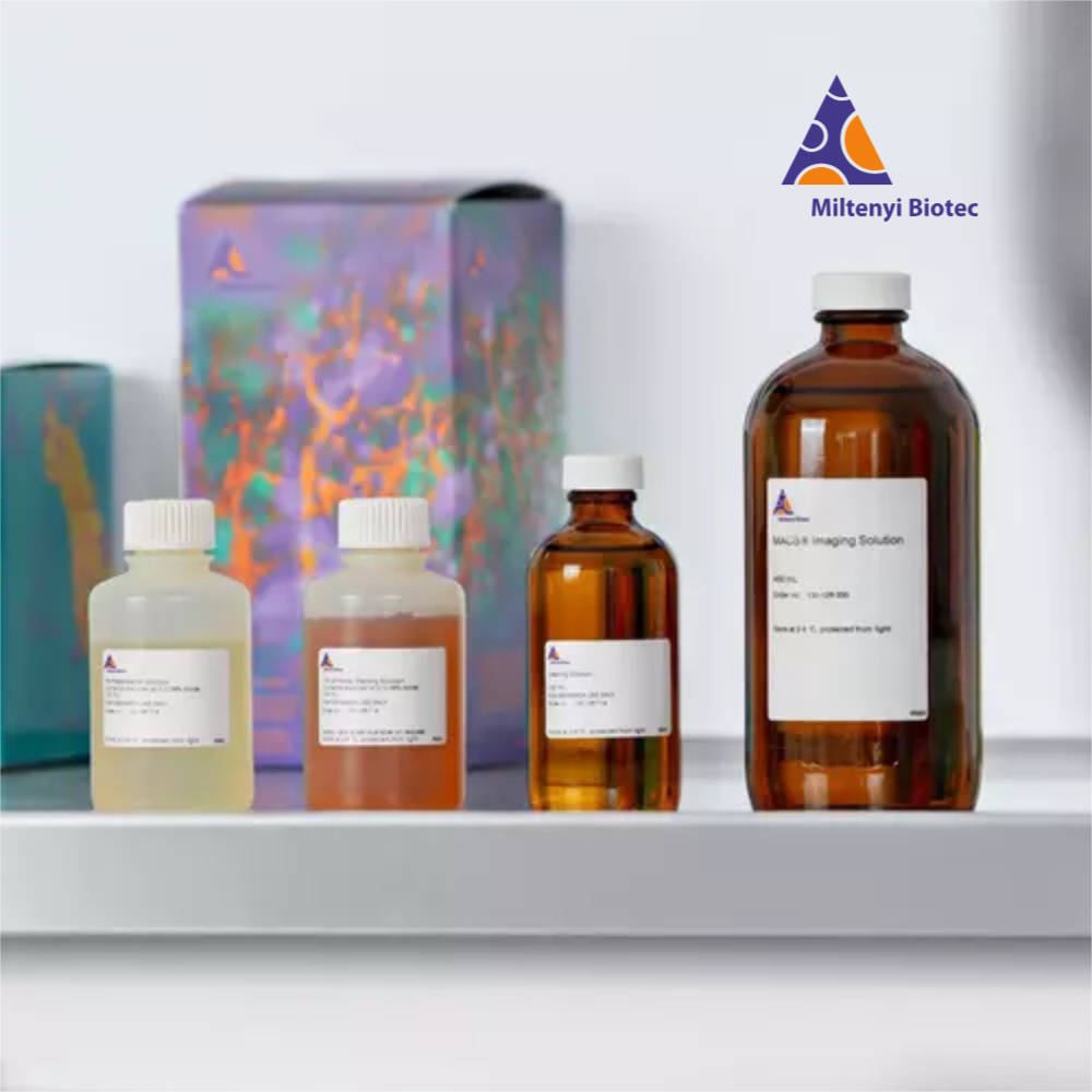


組織透明化試劑
層光顯微鏡三維成像解決方案
專為空間生物學研究設計的高效透明化處理系統,提升樣本透光性與3D影像成像品質

組織透明化背景知識
組織透明化技術是現代生物醫學研究中不可或缺的重要工具,特別適用於"層光顯微鏡"(Light Sheet Microscopy)的三維成像應用。透過有效去除組織中的脂質並調整折射率,此技術能大幅提升樣本的透光性,使研究人員能夠深入觀察完整組織的內部結構。
MACS® 組織透明化系列產品採用溶劑基透明化方法,能夠有效處理從小型器官樣本到大體積腦組織的各種生物樣本。此技術在"空間生物學"、"神經科學"研究以及"藥物研發"領域具有廣泛應用前景。
保護內生性訊號的溶劑基透明化處理
為了在溶劑基透明化過程中保護珍貴的內生性訊號(如GFP訊號),需要對標準方法進行輕微調整。脫水步驟應使用精確調節pH值的叔丁醇(tertiary butanol),而非乙醇。此調整方式適用於MACS Clearing Kit。

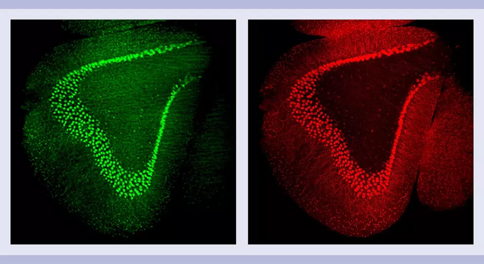
圖說:使用MACS Clearing Kit專用方案透明化處理的GAD67-EGFP小鼠腦部。保護的內生性GFP訊號(左,綠色)對比 GFP抗體(Vio R667)染色(右,紅色)。
* 備注:對於這些組織,可使用與MACS Clearing Kit相同的方案。更多相容性與操作說明請參閱使用說明。
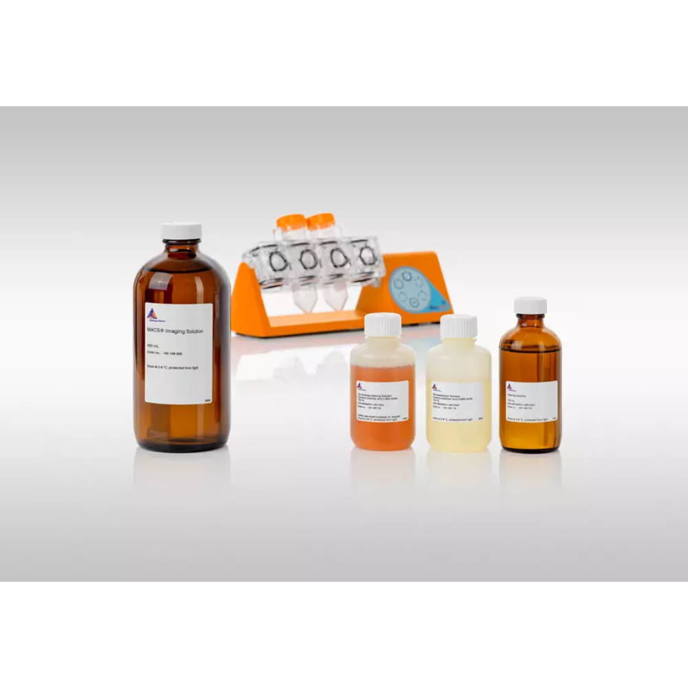
產品套件比較

MACS® Deep Clearing Kit
專為大體積樣本設計的深度透明化解決方案,適用於完整小鼠腦部等大型組織的三維成像分析。
- 適用於完整小鼠腦部處理
- 大體積樣本透明化
- 高效能透明化處理
- 專業級研究應用
- Order no. 130-136-470

MACS® Clearing Kit
通用型組織透明化套件,適用於多種組織類型,包含內生性螢光蛋白訊號保護功能。
- 多種組織類型適用
- 保護內生性訊號
- 腫瘤與異種移植研究
- 類器官與球狀體處理
- Order no. 130-126-719
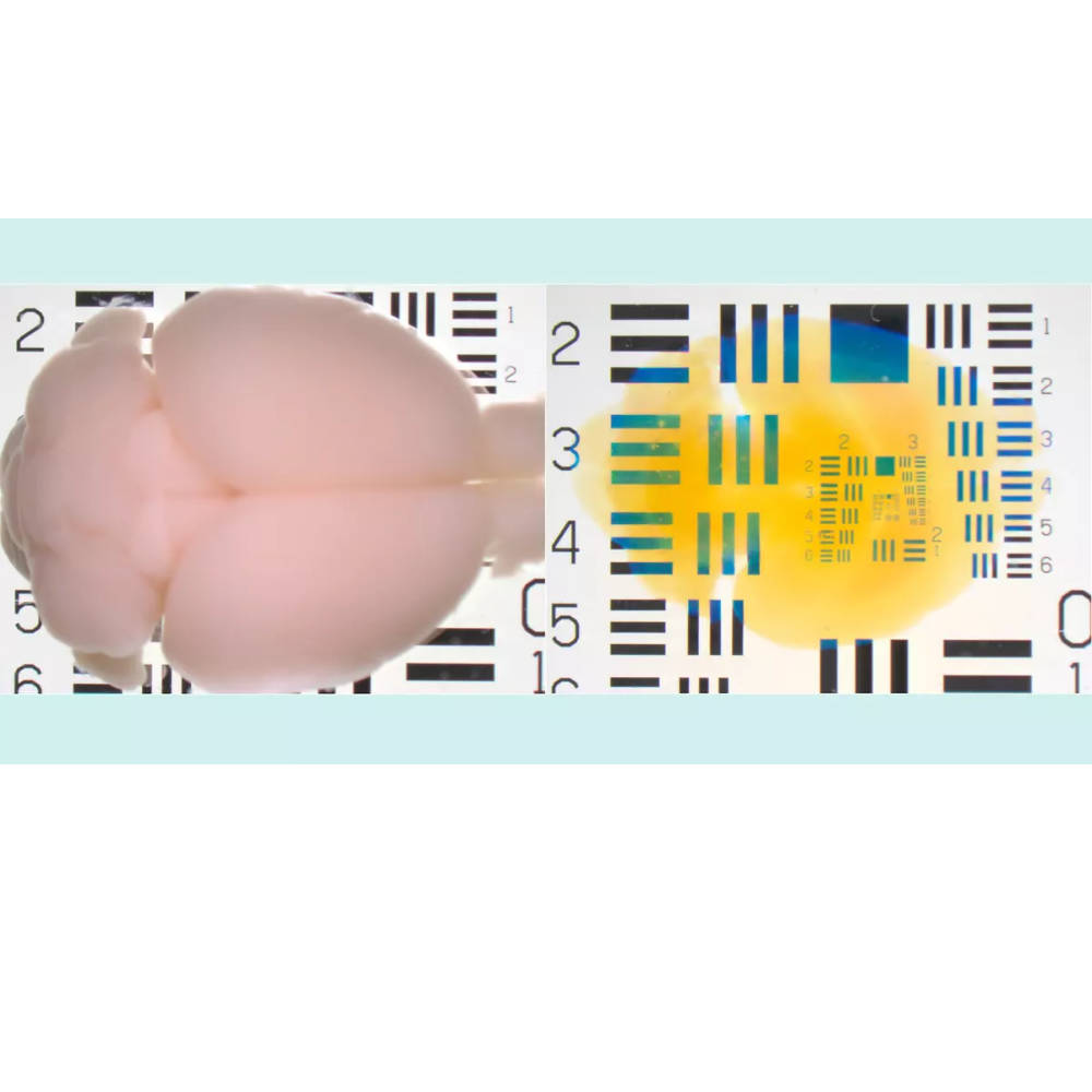
透明化效果展示


使用方案與適用範圍
| Protocol | MACS Deep Clearing Kit | MACS Clearing Kit |
|---|---|---|
| Mouse whole brain | Available | |
| Mouse brain hemisphere | Available | Available |
| Mouse brain hemispheres with preservation of endogenous fluorescent protein signal | * | Available |
| Human (blood-rich) tumors and xenografts | * | Available |
| Organoids and spheroids | * | Available |
| Organoids and spheroids (high-throughput) | * | Available |
| Mouse spleen, kidney, and liver | Under development | Available |
| Mouse lung and intestine | Under development | Available |
| Mouse lymph nodes | * | Available |
| Mouse embryo | Under development | Available |
* 這些組織可套用與 MACS Clearing Kit 相同的操作流程。更多相容性資訊與操作指引,請參閱相關實驗流程。
* Tissue clearing and immunolabeling protocols
* 點選 Available 可連線到Miltenyi觀看Protocol, 若出現404 請通知博克人員
技術要點與實用技巧
組織透明化成功要點
- 樣本前處理:確保組織固定充分,避免過度固定影響透明化效果
- 溫度控制:維持適當的處理溫度,確保試劑效能最佳化
- 時間管控:依據組織大小調整處理時間,避免過度處理
- 溶劑品質:使用高品質溶劑,確保透明化結果一致性
- 儲存條件:透明化後樣本需妥善保存,維持成像品質
常見問題
Technical Documentation & Advanced Applications
Note: The following technical overview is provided in English for international research community reference and SEO enhancement.
Advanced Tissue Clearing Reagents for Light Sheet Microscopy: Revolutionizing 3D Biomedical Imaging and Spatial Biology Research
In the rapidly evolving landscape of biomedical research, tissue clearing technology has emerged as an indispensable tool for advancing our understanding of complex biological systems. This revolutionary approach enables researchers to achieve unprecedented visualization of intact tissue specimens through light sheet microscopy, opening new frontiers in spatial biology, neuroscience, and drug development research.
Understanding Tissue Clearing Technology
Tissue clearing represents a transformative methodology that addresses one of the fundamental challenges in biological imaging: the optical opacity of biological tissues. Traditional histological approaches require tissue sectioning, which inevitably destroys the three-dimensional architecture that is crucial for understanding cellular relationships and tissue organization. Tissue clearing technology overcomes this limitation by rendering biological specimens optically transparent while preserving their structural integrity.
The process involves the systematic removal of light-scattering components, primarily lipids, from biological tissues, followed by refractive index matching to minimize light scattering and absorption. This approach enables deep tissue penetration of light, facilitating high-resolution imaging of intact organs and large tissue specimens that would otherwise be impossible to visualize in their native three-dimensional context.
MACS Tissue Clearing Solutions: Comprehensive Portfolio for Diverse Applications
The MACS tissue clearing reagent portfolio represents a sophisticated approach to biological specimen preparation, offering researchers two distinct but complementary solutions tailored to specific research requirements and sample characteristics.
MACS Deep Clearing Kit: Optimized for Large Volume Specimens
The MACS Deep Clearing Kit addresses the unique challenges associated with large volume specimen processing, particularly whole mouse brain specimens and other substantial tissue samples. This specialized formulation has been engineered to penetrate dense tissue matrices effectively while maintaining consistent clearing performance throughout the entire specimen volume.
The deep clearing methodology employs optimized solvent-based protocols that ensure uniform tissue transparency across specimens that may measure several centimeters in diameter. This capability is particularly valuable for neuroscience research applications where intact brain architecture must be preserved to study neural circuit organization, connectivity patterns, and pathological changes across multiple brain regions simultaneously.
Research applications for the MACS Deep Clearing Kit extend beyond neuroscience to include developmental biology studies requiring whole embryo analysis, cancer research involving large tumor specimens, and comparative anatomy investigations. The kit's robust formulation ensures reproducible results across different tissue types while maintaining compatibility with various fluorescent labeling strategies.
MACS Clearing Kit: Universal Solution with Signal Preservation
The MACS Clearing Kit offers versatility for a broader range of tissue types and experimental approaches. This universal clearing solution has been specifically designed to accommodate diverse specimen sizes and tissue characteristics while providing exceptional preservation of endogenous fluorescent signals.
One of the most significant advantages of the MACS Clearing Kit is its ability to protect endogenous fluorescent protein signals, such as GFP, during the clearing process. Traditional clearing protocols often compromise fluorescent protein integrity due to harsh chemical treatments or extreme pH conditions. The MACS Clearing Kit addresses this challenge through carefully optimized pH buffering and the use of tertiary butanol for dehydration steps, replacing conventional ethanol-based approaches that can denature fluorescent proteins.
This signal preservation capability is particularly valuable for research involving transgenic animal models, where endogenous fluorescent reporters provide crucial information about cell identity, gene expression patterns, and cellular dynamics. The ability to maintain these signals while achieving tissue transparency enables researchers to conduct comprehensive spatial analysis without sacrificing molecular information.
Protocol Diversity and Application Scope
The MACS tissue clearing system supports an extensive range of biological specimens, reflecting the diverse needs of contemporary biomedical research. Protocol availability spans multiple tissue types and experimental contexts, ensuring researchers can access optimized procedures for their specific applications.
Neurobiological applications include protocols for whole mouse brain processing, brain hemisphere analysis, and specialized procedures for preserving endogenous fluorescent protein signals in neural tissues. These protocols have been validated across multiple research contexts and provide reliable results for studies investigating neural development, neurodegenerative diseases, and brain connectivity patterns.
Cancer research applications encompass protocols for human tumor specimens and patient-derived xenografts, including blood-rich tissues that present particular challenges for clearing procedures. These specialized protocols address the unique optical properties of tumor tissues while maintaining the ability to visualize cancer cell distribution, vascular architecture, and immune cell infiltration patterns.
The system also supports organoid and spheroid applications, including high-throughput approaches using specialized sample holders. This capability is increasingly important as organoid-based research models gain prominence in drug discovery, disease modeling, and regenerative medicine applications.
Additional protocol support extends to various organ systems, including liver, spleen, kidney, lung, intestine, lymph nodes, and embryonic tissues. This comprehensive coverage ensures researchers across multiple disciplines can leverage tissue clearing technology for their investigations.
Technical Advantages and Performance Characteristics
The MACS tissue clearing system incorporates several technical innovations that distinguish it from alternative clearing approaches. The solvent-based methodology provides superior lipid removal efficiency while maintaining tissue morphology and protein accessibility for immunolabeling procedures.
Temperature control protocols ensure optimal clearing kinetics while preventing tissue damage or protein denaturation. Processing times are optimized for different tissue sizes, with small specimens typically requiring several hours to complete clearing, while large volume samples may require several days for optimal transparency.
Quality control measures ensure consistent results across different tissue batches and experimental conditions. The reagent formulations maintain stability under standard laboratory storage conditions, providing researchers with reliable performance over extended experimental timelines.
Safety considerations have been incorporated throughout the system design, with reagent formulations selected to minimize health hazards while maintaining clearing efficacy. Standard laboratory safety protocols are sufficient for safe handling and use of the clearing reagents.
Integration with Advanced Imaging Systems
The MACS tissue clearing system has been specifically designed for compatibility with light sheet microscopy platforms, including the UltraMicroscope series. This integration ensures optimal imaging performance and simplified workflow implementation.
Light sheet microscopy represents the ideal imaging modality for cleared specimens due to its ability to provide rapid, high-resolution imaging of large specimens with minimal photobleaching and phototoxicity. The optical characteristics of cleared tissues are well-matched to light sheet illumination geometry, enabling deep tissue penetration and uniform illumination across specimen volumes.
Advanced imaging capabilities include multi-channel fluorescence acquisition, enabling simultaneous visualization of multiple molecular targets within the same specimen. This multiplexing capability is essential for comprehensive spatial biology investigations that require correlation of multiple biological parameters.
Research Impact and Future Directions
Tissue clearing technology is driving significant advances across multiple research domains. In neuroscience, cleared brain specimens are providing unprecedented insights into neural circuit organization, revealing connectivity patterns that were previously inaccessible through conventional approaches. These discoveries are advancing our understanding of brain development, function, and disease mechanisms.
Cancer research applications are yielding new insights into tumor heterogeneity, metastatic processes, and therapeutic response mechanisms. The ability to visualize intact tumor architecture while maintaining cellular and molecular resolution is transforming cancer biology research and informing new therapeutic strategies.
Developmental biology investigations are benefiting from the ability to trace cellular lineages and morphogenetic processes throughout entire embryos, providing comprehensive understanding of developmental mechanisms that govern organ formation and tissue patterning.
Conclusion
The MACS tissue clearing reagent system represents a sophisticated solution for modern biomedical research requiring three-dimensional tissue analysis. Through careful optimization of clearing protocols, signal preservation capabilities, and broad specimen compatibility, this system enables researchers to unlock new insights into biological systems that were previously inaccessible.
As spatial biology continues to emerge as a critical research frontier, tissue clearing technology will play an increasingly important role in advancing our understanding of complex biological processes. The combination of advanced clearing reagents, optimized protocols, and compatible imaging systems provides researchers with the tools necessary to address fundamental questions in biology and medicine while maintaining the highest standards of experimental rigor and reproducibility.
The future of tissue clearing technology promises continued innovation in reagent formulations, protocol optimization, and imaging integration, ensuring that researchers will have access to increasingly powerful tools for spatial biology investigations. This technological evolution will undoubtedly contribute to breakthrough discoveries that advance human health and deepen our understanding of life sciences.