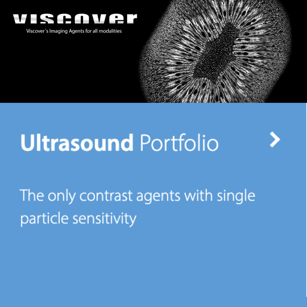
實驗動物超音波顯影劑專家
Viscover PolySon™ 系列 - 唯一具備單顆粒靈敏度的硬殼微氣泡顯影劑
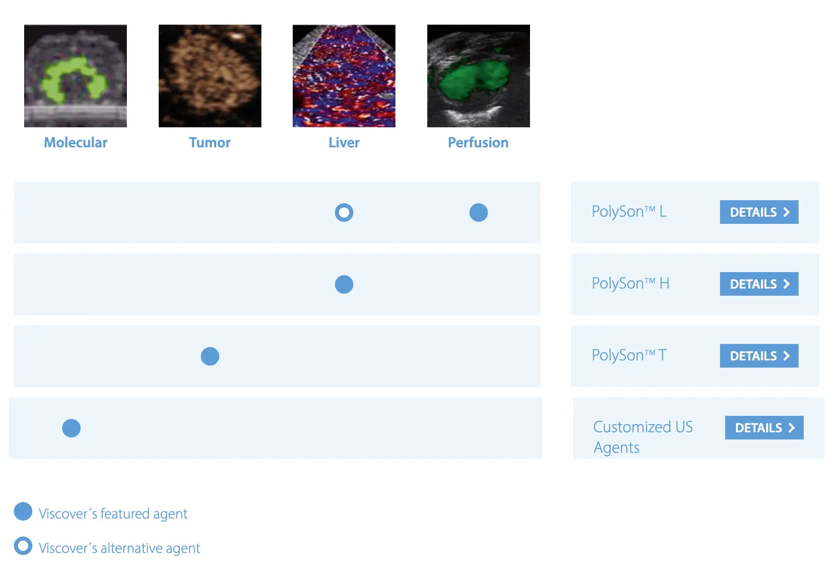
PolySon™ 超音波顯影劑產品系列
獨特的硬殼微氣泡技術
Viscover PolySon™ L、PolySon™ H 和 PolySon™ T 超音波顯影劑是市場上唯一的硬殼超音波微氣泡 (Hard-shell Ultrasound Microbubbles),具備理想的生理特性和對比特性,適用於所有典型的超音波檢查方法。
高回聲性與單顆粒靈敏度
PolySon 顯影劑是微米級的充氣聚合物顆粒或微氣泡 (Microbubbles)。由穩定的惰性聚合物外殼 (Inert Polymeric Shell) 組成,展現可調節的聲學特性 (Tunable Acoustic Properties),能實現快速可靠的超音波成像。此外,PolySon 顯影劑還提供體內單氣泡量化 (In Vivo Single Bubble Quantification) 的額外功能,透過「氣泡破壞」方法 (Bubble Destructive Methods),即刺激聲發射 (Stimulated Acoustic Emission, SAE) 技術。
專為高要求的臨床前應用開發
PolySon™ L
硬殼超音波微氣泡顯影劑,最適合器官灌注測量 (Organ Perfusion Measurements) 以及肝臟和脾臟的可視化 (Visualization of Liver and Spleen)
PolySon™ H
提供與 PolySon™ L 相同的功能,但針對體表高聲壓測量 (Measurements at Body Surface with High Acoustic Pressures) 進行優化
PolySon™ T
硬殼超音波微氣泡顯影劑,專為腫瘤成像 (Imaging of Tumors) 優化設計
應用範圍 (Application Areas)
- 器官灌注評估 (Organ Perfusion Assessment) - 心血管系統、腎臟系統、腫瘤內血管灌注研究
- 肝臟與脾臟成像 (Liver and Spleen Imaging) - 網狀內皮系統 (RES) 攝取觀察
- 腫瘤可視化 (Tumor Visualization) - 血管化腫瘤的增強對比成像
- 敏感顆粒聲學量化 (Sensitive Particle Acoustic Quantification, SPAQ) - 高濃度顯影劑定量分析
- 心臟灌注成像 (Cardiac Perfusion Imaging) - 心肌對比超音波評估
- 分子影像研究 (Molecular Imaging Research) - 標靶特異性超音波造影
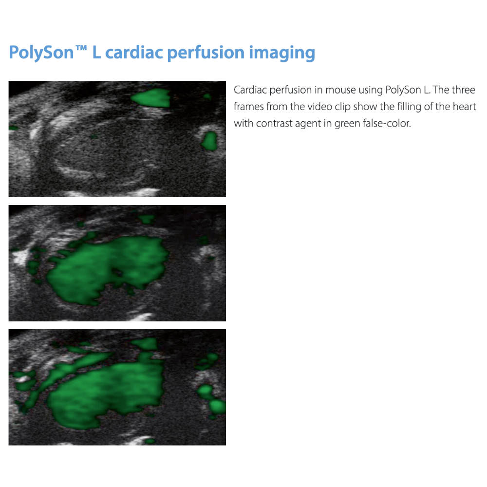
PolySon™ L 超音波顯影劑
Optimized for Low Acoustic Pressure Perfusion Imaging
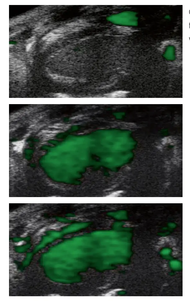
小鼠心臟灌注成像使用 PolySon™ L 顯影劑 - 綠色偽彩顯示心臟填充過程
產品編號 (Order Number)
| 產品規格 | 訂購編號 |
|---|---|
| PolySon™ L 超音波顯影劑 (1 × 5 次注射) | 130-095-150 |
| PolySon™ L 超音波顯影劑 (5 × 5 次注射) | 130-095-151 |
產品特性 (Product Features)
- 高回聲性 (Highly Echogenic) - 在所有常用頻率下提供最佳對比度
- 微觀解析度 (Microscopic Resolutions) - 能夠在顯微級解析度下可視化器官灌注
- 低聲壓優化 (Low Acoustic Pressure Optimization) - Viscover 領先的灌注成像超音波顯影劑
- SAE 成像能力 (SAE Imaging Capability) - 使用低能量超音波束 (Low Mechanical Index) 執行刺激聲發射成像
理化特性 (Physico-chemical Properties)
- 顆粒大小 (Particle Size, Number-weighted): 1-3 µm
- 組成結構 (Composition): 微米級空氣填充聚合物顆粒 (Micrometer-sized Air-filled Polymeric Particles)
- 外殼類型 (Shell Type): 穩定惰性聚合物硬殼 (Stable Inert Polymeric Hard Shell)
- 循環時間 (Circulation Time): 靜脈注射後在血液中循環長達 10 分鐘
主要應用 (Primary Applications)
- 灌注模式評估 (Evaluation of Perfusion Patterns) - 心血管系統、腎臟系統灌注研究
- 心臟灌注成像 (Cardiac Perfusion Imaging) - 心肌對比超音波心動圖 (Myocardial Contrast Echocardiography)
- 肝臟與脾臟可視化 (Liver and Spleen Visualization) - 網狀內皮系統 (RES) 攝取觀察
- 腫瘤內血管灌注 (Intratumoral Vessel Perfusion) - 腫瘤血管化評估
適用技術 (Compatible Techniques)
諧波成像 (Harmonic Imaging) 次諧波成像 (Subharmonic Imaging) B 模式成像 (B-mode Imaging) 刺激聲發射 (SAE - Low MI) 敏感顆粒聲學量化 (SPAQ)
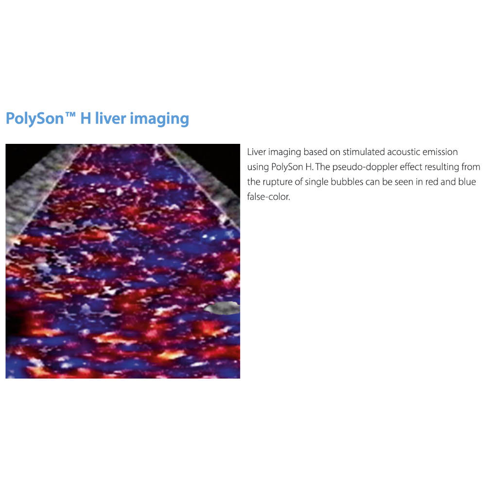
PolySon™ H 超音波顯影劑
Optimized for High Acoustic Pressure Liver Imaging
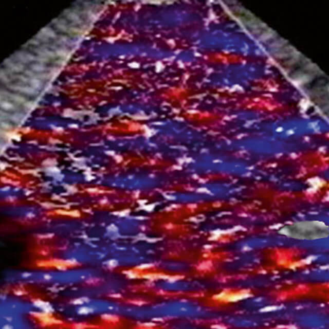
使用 PolySon™ H 進行肝臟刺激聲發射成像 - 紅藍偽彩顯示單氣泡破裂產生的偽都卜勒效應
產品編號 (Order Number)
| 產品規格 | 訂購編號 |
|---|---|
| PolySon™ H 超音波顯影劑 (1 × 5 次注射) | 130-095-152 |
| PolySon™ H 超音波顯影劑 (5 × 5 次注射) | 130-095-153 |
產品特性 (Product Features)
- 高度堅固微氣泡 (Highly Robust Microbubbles) - 專為高聲壓肝臟成像優化
- 高能量 SAE 成像 (High-energy SAE Imaging) - 完美適用於基於可誘導氣泡破裂的刺激聲發射成像
- 體表高壓測量 (High Acoustic Pressure Surface Measurements) - 提供與 PolySon™ L 相同的功能,但針對高機械指數優化
- 偽都卜勒效應 (Pseudo-Doppler Effect) - 單氣泡破裂產生的獨特聲學信號
理化特性 (Physico-chemical Properties)
- 組成結構 (Composition): 高強度聚合物硬殼微氣泡 (High-strength Polymeric Hard-shell Microbubbles)
- 耐壓特性 (Pressure Resistance): 優化用於高機械指數 (High Mechanical Index) 超音波
- 外殼穩定性 (Shell Stability): 增強型惰性聚合物外殼,可承受高能量超音波束
- 循環時間 (Circulation Time): 靜脈注射後在血液中循環長達 10 分鐘
主要應用 (Primary Applications)
- 高壓肝臟成像 (High-pressure Liver Imaging) - 體表高聲壓條件下的肝實質組織評估
- 脾臟實質評估 (Splenic Parenchyma Evaluation) - 組織特異性超音波造影
- 高機械指數 SAE (High Mechanical Index SAE) - 使用高能量超音波束的刺激聲發射成像
- 肝腫瘤成像 (Liver Tumor Imaging) - 肝臟病變的增強對比可視化
適用技術 (Compatible Techniques)
諧波成像 (Harmonic Imaging) 次諧波成像 (Subharmonic Imaging) B 模式成像 (B-mode Imaging) 刺激聲發射 (SAE - High MI) 敏感顆粒聲學量化 (SPAQ) 體表高頻超音波 (Surface High-frequency US)
技術優勢 (Technical Advantages)
PolySon™ H 是市場上唯一能夠在高機械指數條件下保持穩定性的硬殼微氣泡顯影劑。其獨特的偽都卜勒效應 (Pseudo-Doppler Shifts) 源於非移動微氣泡的破壞過程,為組織特異性超音波造影提供了革命性的技術突破。特別適合需要高能量超音波束的體表成像和深層組織評估。
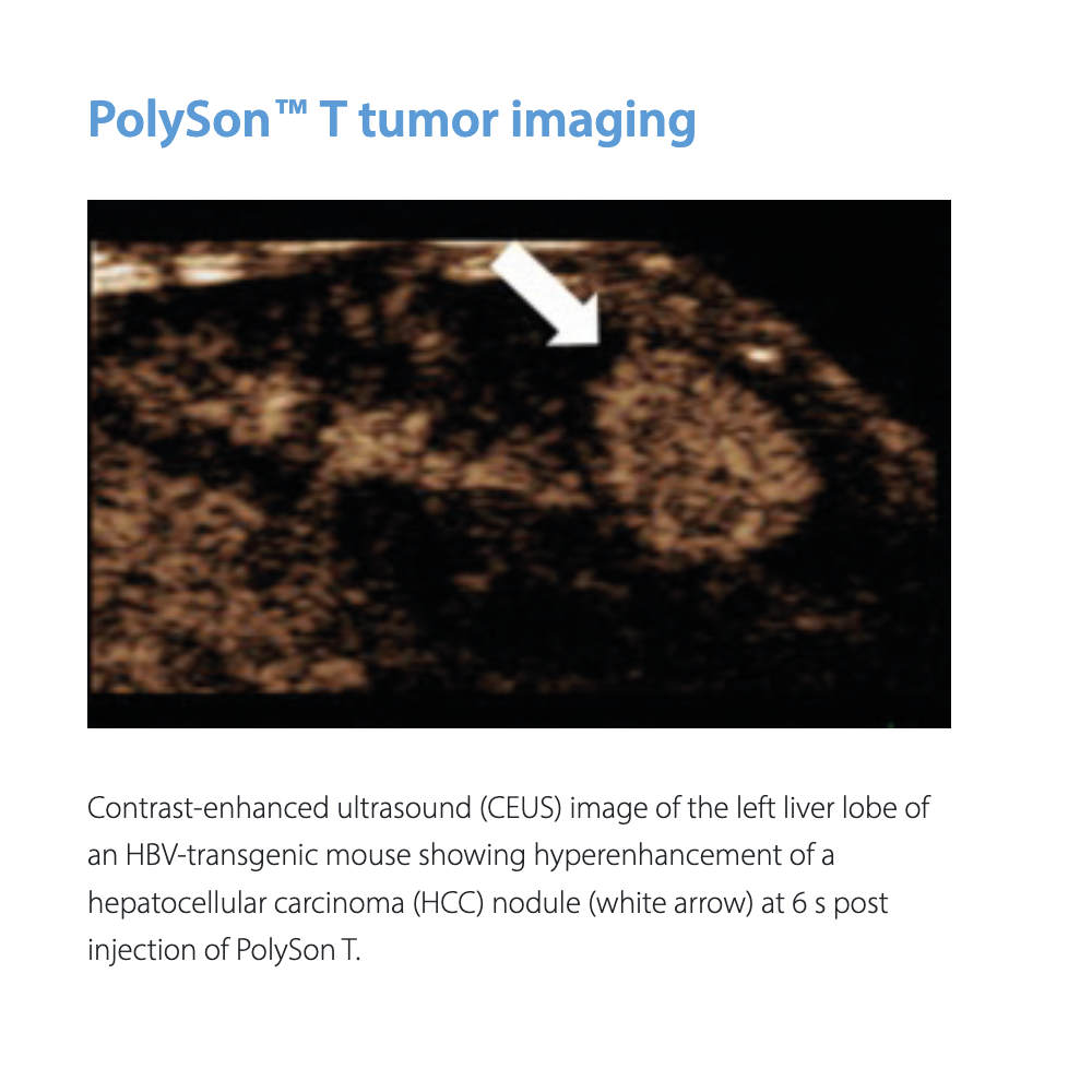
PolySon™ T 超音波顯影劑
Optimized for Tumor Imaging with Extended Blood Half-life
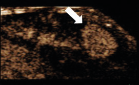
HBV 轉基因小鼠肝細胞癌 (HCC) 結節的對比增強超音波 (CEUS) 成像 - 注射 PolySon™ T 後 6 秒
產品編號 (Order Number)
| 產品規格 | 訂購編號 |
|---|---|
| PolySon™ T 超音波顯影劑 (1 × 5 次注射) | 130-095-148 |
| PolySon™ T 超音波顯影劑 (5 × 5 次注射) | 130-095-149 |
產品特性 (Product Features)
- 極高對比增強 (Exceptionally High Contrast Enhancement) - 腫瘤成像優化配方
- 延長血半衰期 (Long Blood Half-life) - 提供更長的成像時間窗口
- 血管化腫瘤可視化 (Vascularized Tumor Visualization) - 改善動物腫瘤模型中的腫瘤觀察能力
- 增強灌注評估 (Enhanced Perfusion Assessment) - 腫瘤血管化程度的精確評估
理化特性 (Physico-chemical Properties)
- 組成結構 (Composition): 專為腫瘤成像優化的聚合物微氣泡 (Tumor-optimized Polymeric Microbubbles)
- 血液循環 (Blood Circulation): 延長的血半衰期提供持續的對比增強
- 對比性能 (Contrast Performance): 在腫瘤組織中展現卓越的回聲增強
- 血管親和性 (Vascular Affinity): 優化的血管滯留特性
主要應用 (Primary Applications)
- 腫瘤對比增強超音波 (Tumor Contrast-Enhanced Ultrasound, CEUS) - 血管化腫瘤的高對比成像
- 肝細胞癌評估 (Hepatocellular Carcinoma Assessment) - HCC 結節的超增強 (Hyperenhancement) 可視化
- 動物腫瘤模型研究 (Animal Tumor Model Studies) - 實驗性腫瘤的生長監測和治療反應評估
- 腫瘤血管化評估 (Tumor Vasculature Assessment) - 腫瘤新生血管 (Tumor Angiogenesis) 的定量分析
- 抗癌療效監測 (Anti-cancer Therapy Monitoring) - 治療後腫瘤灌注變化的追蹤
適用技術 (Compatible Techniques)
對比增強超音波 (CEUS) 諧波成像 (Harmonic Imaging) 功率都卜勒 (Power Doppler) B 模式成像 (B-mode Imaging) 腫瘤灌注量化 (Tumor Perfusion Quantification)
技術優勢 (Technical Advantages)
PolySon™ T 是專為癌症研究 (Cancer Research) 設計的突破性超音波顯影劑。其獨特的配方結合了極高的對比增強能力和延長的血半衰期,使研究人員能夠在更長的時間窗口內評估腫瘤的血管化程度和灌注模式。特別適用於轉基因腫瘤模型 (Transgenic Tumor Models)、皮下異種移植 (Subcutaneous Xenografts) 和原位腫瘤模型 (Orthotopic Tumor Models) 的超音波監測。
臨床前研究應用案例 (Preclinical Research Applications)
- HCC 動物模型 (HCC Animal Models) - HBV 轉基因小鼠的肝細胞癌結節可視化
- 抗血管新生療法評估 (Anti-angiogenic Therapy Evaluation) - 治療前後腫瘤血管變化監測
- 藥物遞送研究 (Drug Delivery Studies) - 腫瘤靶向治療的灌注效果評估
- 轉移性腫瘤檢測 (Metastatic Tumor Detection) - 肝臟和其他器官的微小轉移灶識別
Viscover™ 客製化顯影劑服務
Customized Imaging Agents for Specific Research Needs
量身訂製的解決方案
除了標準的 PolySon™ 系列產品,Viscover 還提供客製化超音波顯影劑開發服務 (Customized Ultrasound Contrast Agent Development)。我們的專業團隊可以根據您特定的研究需求,設計和製造符合您實驗要求的專屬顯影劑。
客製化服務內容 (Customization Services)
- 粒徑調整 (Particle Size Adjustment) - 根據目標器官和超音波頻率優化微氣泡大小
- 外殼改質 (Shell Modification) - 調整聚合物組成以改變聲學特性和循環時間
- 表面功能化 (Surface Functionalization) - 添加標靶配體 (Targeting Ligands) 用於分子影像研究
- 藥物負載 (Drug Loading) - 開發具有治療功能的微氣泡平台
- 多模態成像 (Multimodal Imaging) - 結合螢光或 MRI 對比劑的複合顯影劑
應用領域 (Application Areas)
分子影像 (Molecular Imaging)
標靶特異性微氣泡用於生物標記檢測和疾病早期診斷
治療性超音波 (Therapeutic Ultrasound)
藥物遞送微氣泡和超音波介導的基因轉染
血腦屏障開放 (BBB Opening)
聚焦超音波結合微氣泡的神經科學應用
特殊器官成像 (Specialized Organ Imaging)
針對特定器官或組織類型優化的顯影劑
聯繫我們
如果您有特殊的研究需求或希望開發客製化的超音波顯影劑,歡迎聯繫博克科技獲取更多資訊和技術支援。我們的專業團隊將協助您選擇最適合的產品或設計專屬的解決方案。
超音波影像範例
Data Gallery - Ultrasound Imaging Examples

Viscover 提供豐富的應用範例和影像資料庫,展示 PolySon™ 系列顯影劑在各種實驗動物模型中的實際應用效果。這些範例涵蓋了心臟灌注、肝臟成像、腫瘤可視化、腎臟灌注等多個研究領域。
影像資料庫內容 (Data Gallery Contents)
- 心血管系統成像 (Cardiovascular Imaging) - 心肌灌注、心室功能、血管造影
- 腹部器官成像 (Abdominal Organ Imaging) - 肝臟、脾臟、腎臟的對比增強影像
- 腫瘤學應用 (Oncology Applications) - 各類腫瘤模型的 CEUS 影像
- SAE 技術範例 (SAE Technique Examples) - 刺激聲發射成像的偽都卜勒效應展示
- 灌注量化分析 (Perfusion Quantification Analysis) - SPAQ 方法的定量數據
相關科學文獻
Scientific References and Publications
主要研究論文 (Key Research Publications)
Reinhardt, M. et al. (2005)
Sensitive particle acoustic quantification (SPAQ): a new ultrasound-based approach for the quantification of ultrasound contrast media in high concentrations.
Investigative Radiology 40: 2-7
Tiemann, K. et al. (2000)
Stimulated acoustic emission: pseudo-Doppler shifts seen during the destruction of nonmoving microbubbles.
Ultrasound in Medicine & Biology 26: 1161-1167
Hauff, P. et al. (2004)
Molecular targeting of lymph nodes with L-selectin ligand-specific US contrast agent: a feasibility study in mice and dogs.
Journal of Immunotherapy 231: 667-673
Forsberg, F. et al. (1999)
Tissue-specific US contrast agent for evaluation of hepatic and splenic parenchyma.
Radiology 210: 125-132
Teupe, C. et al. (2001)
Assessment of myocardial perfusion by myocardial contrast echocardiography using harmonic power and the transvenous contrast agent SHU 563A in acute coronary occlusion after reperfusion.
Circulation
常見問題
Frequently Asked Questions
選擇 Viscover PolySon™ 的理由
唯一的硬殼技術
市場上唯一的硬殼聚合物微氣泡,提供無與倫比的聲學穩定性和單顆粒靈敏度
全方位應用覆蓋
從器官灌注到腫瘤成像,從低壓到高壓成像,完整的產品線滿足各種研究需求
先進成像技術
支援 SAE、SPAQ 等前沿超音波技術,開創實驗動物成像的新可能
專業技術支援
完整的技術文獻、應用範例和專業諮詢服務,確保您的研究成功
開始您的超音波研究之旅
無論您是進行心血管研究、腫瘤學研究還是器官灌注評估,Viscover PolySon™ 系列都能為您提供可靠的成像解決方案。立即聯繫博克科技,讓我們的專家協助您選擇最適合的產品,並為您的研究提供專業支援。
English SEO Description
Viscover PolySon™ series represents the gold standard in preclinical ultrasound contrast agents, featuring unique hard-shell polymeric microbubbles specifically engineered for advanced animal imaging research. Our comprehensive portfolio includes three specialized formulations: PolySon™ L for low acoustic pressure perfusion imaging, PolySon™ H for high acoustic pressure surface measurements, and PolySon™ T for optimized tumor visualization.
These innovative hard-shell microbubbles offer unprecedented single particle sensitivity and exceptional echogenicity across all commonly used ultrasound frequencies, including high-frequency microscopy applications above 30 MHz. The stable inert polymeric shell structure enables tunable acoustic properties, making PolySon agents compatible with various ultrasound techniques including harmonic imaging, subharmonic imaging, B-mode imaging, and advanced stimulated acoustic emission (SAE) methods.
PolySon™ L (Order No. 130-095-150/151) is specifically formulated for organ perfusion measurements and cardiac imaging. With a particle size of 1-3 µm, it delivers optimal contrast at microscopic resolutions and excels in myocardial perfusion studies using low mechanical index SAE techniques. The agent circulates in the blood pool for up to 10 minutes post-injection and is taken up by the reticuloendothelial system (RES) in liver and spleen.
PolySon™ H (Order No. 130-095-152/153) features highly robust microbubbles optimized for high acoustic pressure imaging of liver parenchyma. This formulation is perfectly suited for high mechanical index SAE imaging, generating distinctive pseudo-Doppler effects from bubble rupture that enable sensitive particle acoustic quantification (SPAQ). It maintains stability under high-energy ultrasound beams, making it ideal for body surface measurements and deep tissue assessment.
PolySon™ T (Order No. 130-095-148/149) represents a breakthrough in tumor imaging, combining exceptionally high contrast enhancement with an extended blood half-life. This specialized formulation significantly improves visualization of vascularized tumors in animal models, including hepatocellular carcinoma (HCC), subcutaneous xenografts, and orthotopic tumor models. The prolonged circulation time provides an extended imaging window for comprehensive tumor perfusion assessment and treatment monitoring.
All PolySon products are compatible with major preclinical ultrasound platforms including Vevo (VisualSonics), Prospect (S-Sharp), and Sonix (Ultrasonix) systems. They support multiple imaging modalities and enable quantitative perfusion analysis through advanced techniques like SPAQ, making them indispensable tools for cardiovascular research, oncology studies, and organ perfusion investigations in small animal models such as mice, rats, and hamsters.
Viscover also offers customized imaging agent development services for specialized research applications, including surface functionalization for molecular imaging, drug-loaded therapeutic microbubbles, and multimodal contrast agents. Our scientific team provides comprehensive technical support, application guidance, and training to ensure successful implementation in your research program. Contact JNH Technologies for detailed product information, ordering, and technical consultation.