服務專線:0800.898.178 info@jnhtech.com.tw
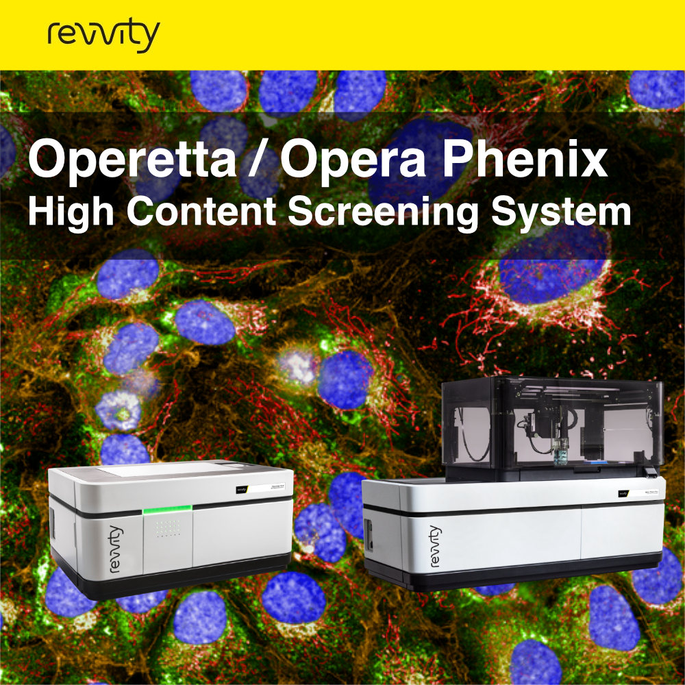
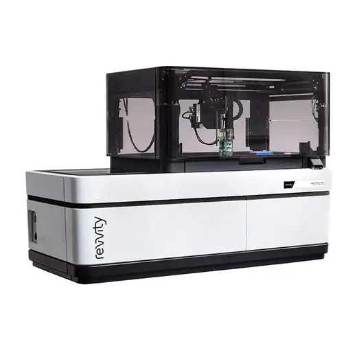
Opera Phenix Plus/ Operetta CLS ™
什麼是高內涵系統?
高內涵細胞成像和分析系統(HCS,High-content screening)是指保持細胞結構完整和功能的情況下,通過自動化成像分析方式,對多孔板中每一個孔的細胞進行單細胞水平的狀態、變化等多參數及總體趨勢的分析。在單一實驗中同步獲取細胞標靶蛋白的空間和時間分佈、表達強度、細胞與胞器的形態和複雜表型、多種細胞亞群的分類等方面的高可靠性的具有統計學意義的分析結果, 同時去除其它細胞與人為誤差的干擾。HCS的結果由圖像分析所得, 兼顧了直觀與批量統計定量的優點,同時輸出單細胞和群體細胞的結果。
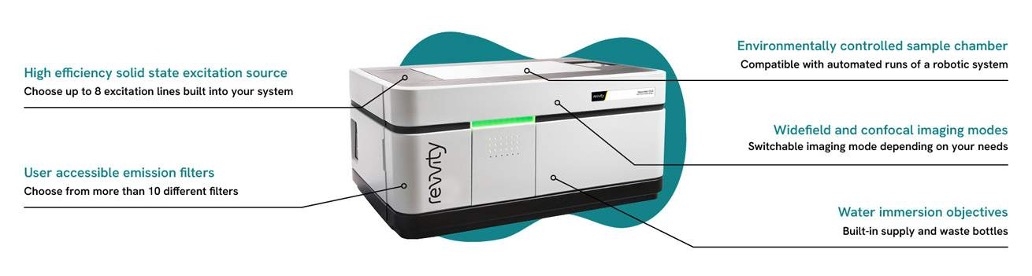
儀器特點
Synchrony專利光路消除交叉干擾
Synchrony光學元件經過專門設計,可以讓雷射更高效率的照射在樣品上,又可捕捉到更多來自樣品的光,無需在速度和靈敏度之間作出取捨。共聚焦的Synchrony光學元件能將交叉干擾降至最低,在不損失靈敏度的前提下最多可以同時採集4條通道,讓您在多色HCS實驗的掃描中達到前所未有的速度。常見顯微成像系統中,同步採集的優勢往往被隨之增強的交叉干擾所掩蓋,這是因為光譜重疊會導致長波長發射的通道檢測到來自短發射波長螢光團的信號。在使用DAPI和Hoechst DNA染料等具有較寬藍色發射譜帶的螢光團時,可能會與綠色螢光蛋白發射譜帶產生重疊,從而降低細胞核轉位等檢測的靈敏度。儘管可以依次採集藍色和綠色通道,但有悖於平行檢測的核心目的。另外,Synchrony Optics™的共焦顯微鏡技術,也可以在解決針孔串擾。因其具有微透鏡增強的雙盤設計,且針孔距離經過優化,適用於像3D微組織這樣的厚樣品,並且雙場激發可在空間和時間上分開相鄰光譜通道的激發。這樣可以實現快速的多通道3D採集,並且實現高影像品質。
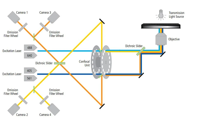
Synchrony 光學元件將相鄰激發線的光路分開,以最大限度地減少同步成像之間的串擾,並最大限度地提高速度和靈敏度。
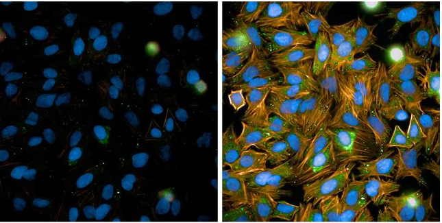
(用Hoechst(DNA)和Alexa488標記的抗微管蛋白抗體染色,再通過傳統同步採集方式成像Hela細胞。Alexa488通道顯示細胞核區域,存在來自Hoechst染料的明顯交叉干擾(左)。而在配備Synchrony光學元件的Opera Phenix系統上進行同樣的染色同步成像時,細胞核中幾乎沒有交叉干擾(右)。)
- 免光纖光路:
在光路耦合設計中,我們創新性的使用免光纖入射技術,以此實現入射光路效率最大化,較之同類產品,激發光入射效率提高30%以上;Operetta的成像系統採用了,自行設計開發的光源和成像光路。特點是具備智慧自我調整光路,使用過程中無需校正。可使成像解析度高及探測器圖元利用率高,並進行精確的Z軸活細胞切片成像和大圖像拼接。
(獨家輪狀光路設計,提供快速激發光源切換,使所有激發光源皆經過相同光路,並減少光路經過的反射鏡)
- 水介質鏡頭可提高解析度:
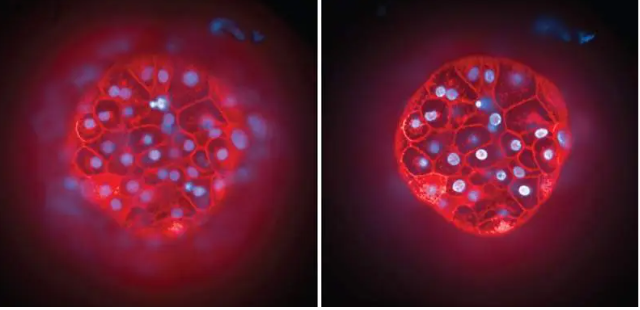
簡稱水鏡,可以自動供水。水鏡可以同時滿足高解析度及高通量掃描的要求,也可以進行DNA損傷、有絲分裂、酵母細胞內質網結構、線粒體形態等微小結構的高清晰度觀察。所有物鏡採用條碼自動識別技術,無需人工設置,可同時裝載6個物鏡,所有水鏡可以同時安裝在儀器上,無需使用者自行更換,可選配水鏡數值孔徑不小於1.0,對比傳統空氣鏡頭,解析度和螢光通透率都有了飛越的提升。

(來自 InSphero 的 3D InSight™ 人類肝臟微組織,用 Hoechst(細胞核,藍色)和 CellMask™ 深紅色質膜染色劑(膜,紅色)標記。左:一般物鏡。右:水鏡。)
- 智慧影像擷取:
HCS 智慧採集技術,可以更準確地定位感興趣的對象,從而顯著縮短採集和分析時間。此功能可透過 Harmony 高內涵分析軟體搭配 PreciScan 外掛程式實現。它實現了低倍率預掃描、影像分析和高倍率重新掃描的完全自動化和整合工作流程。例如,此工作流程可用於使用 5 倍明場,掃描定位 96 孔板所有孔中的 3D 微組織,然後在組織中心以 40 倍放大倍率取得多色螢光 z 堆疊,它也可用於定位共培養中的幹細胞集落或識別大型細胞群中的罕見事件。您可以透過 10 倍預掃描來識別有絲分裂細胞,並透過細胞核的 63 倍成像進行追蹤。 PreciScan 也能夠對斑馬魚或組織切片等較大物體進行高效平鋪成像。
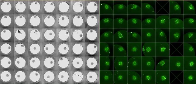
(左圖:使用 PreciScan 軟體以低放大倍率 (10 倍) 進行預掃描,以識別微組織生長的孔。右圖:僅以更高的放大倍率 (20 倍) 重新掃描微組織已生長且微組織位於影像中心的孔, 3D InSight Microtissue courtesy of InSphero AG)
軟體特色
- 標配30多種軟體分析模版:
Harmony™為研究人員提供了30多種得到藥篩人員驗證、SCI雜誌認可的專業且易於使用的分析模塊。以細胞週期研究為例,其中“有絲分裂指數”和“核分類-DNA含量”方案,即可作為細胞凋亡分析的“核斷裂”方案。研究者可以使用現成的分析方法,或者通過簡單積木堆疊構建新的分析方法。
- Preci-scan智慧預掃功能:
利用 PreciScan 智慧影像擷取的強大功能,實現更有效率的高內涵成像和分析。系統可以進行全自動低倍鏡全孔預掃描獲得全景圖,然後自動篩選目標拍攝區域,接下來自動切換到合適的高倍鏡下對目的地區域進行精細掃描,大大縮短拍攝時間、減小資料儲存記憶體,提升實驗效率。此工作流程可用於定位 96 孔板的所有孔中的 3D 微組織,然後在組織中心取得 40 倍率的 z 堆疊圖像,其他應用包括為罕見事件研究定位罕見訊號、高解析度掃描和組織切片等大樣本掃描。從而顯著減少採集和分析時間,這對於 3D 微組織和罕見事件研究特別有價值。
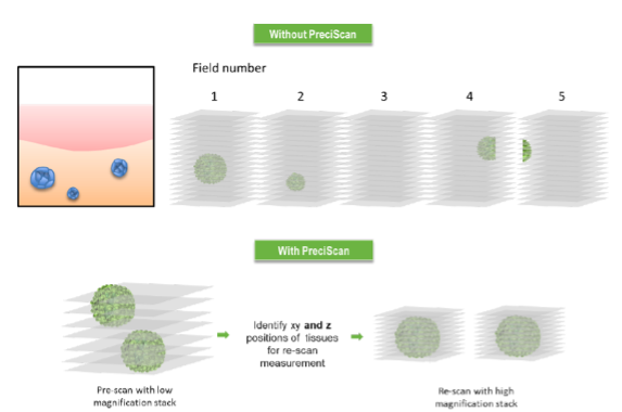
(若無 PreciScan,每個區域需要進行大量的 z 堆疊掃描來覆蓋所有球形體。有PreciScan 的話能在 PreScan 實驗中確定三維物體的 x、y 和 z 坐標,並在 ReScan 實驗中使用這些坐標進行基於物體的成像,從而使物體在 XYZ 空間中居中。
- PhenoLOGIC機器自學習功能:
使用者可通過簡單的圈選-訓練-識別,教導軟體辨別不同的細胞類群或區域,創建自訂的分析演算法。同時可區分多至6種表型,推薦最優區分參數。可自行學習細胞大小、形態、細胞紋理、亞細胞結構,組織形態 結構,信號分佈差參數。
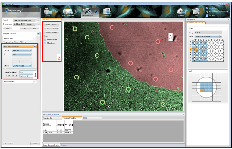
(圖上顯示了機器學習方法的應用。HT1080 細胞從圖像左下角的封閉細胞層遷移到左上角的空白區域。顏色疊加顯示了分割結果(細胞覆蓋區域—綠色,細胞空白區域—紅色)。紅色和綠色圓圈表示用於訓練 Find Texture Region 組件的訓練樣本。1 – 使用 PhenoLOGIC 機器學習的組件。2 – 訓練控制元素。3 – 訓練樣本。)
- Texture 纹理分析功能:
擺脫螢光強度的束縛,根據研究者判讀的經驗進行亞細胞結構的細胞紋理量化分析:根據細胞紋理的不同,將細胞紋理分為Ridge,Valley,Saddle,Edge等多種形態模型,綜合紋理的尺寸,角度,相互關係,量化圖像獲得細胞生物學數據結果。
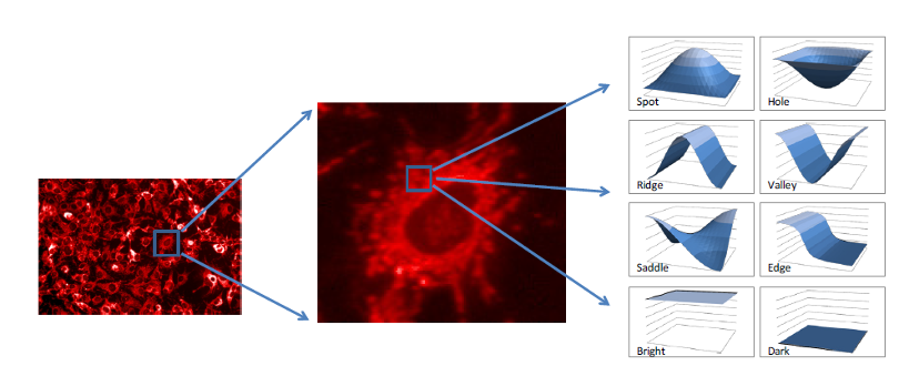
(對於一個區域(此處為圍繞細胞核的細胞質區域)的強度結構進行紋理分析,以查找典型的強度模式,例如“邊緣”、“脊”或“斑點”。生物狀態通過特定特徵或特徵組合的頻率來表達。
- 專利 STAR 窮舉法分析功能:
由軟體對圖像進行全面自主分析,無需任何人工干預,為研究者找到最合適的分析方法,獲得形態學參數不低於207個。STAR專利演算法可用於未知作用機理或未知表型研究中,解決肉眼無法區分細胞表型差異、設置有效的區分參數而導致錯篩和漏篩的難題。
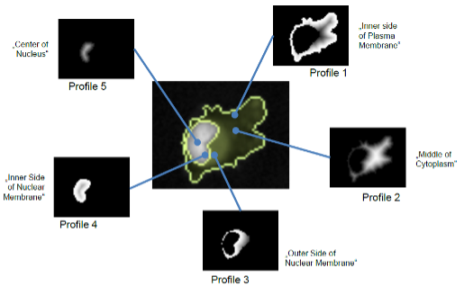
(五個屬性決定了螢光強度相對於細胞(或其他物體)內五個不同預定義區域的位置。顏色顯示了強度的加權情況。白色表示強度的權重較高,黑色表示權重為零。)
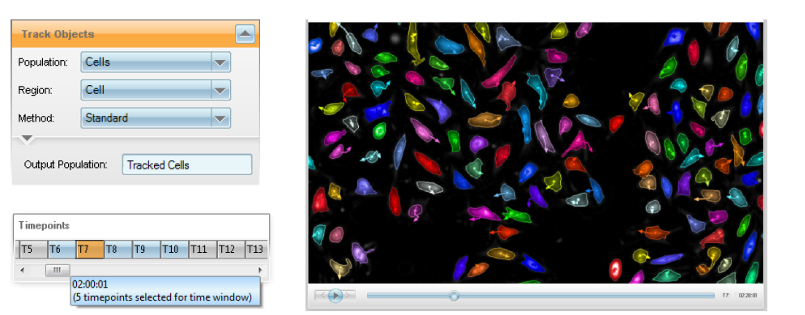
- 動態形態學追蹤功能:
對全部或部分感興趣細胞群進行移動距離、速度、分離、融合、單方向位移、分裂代次等多參數分析,即使無螢光標記細胞也可以輕鬆獲取定量參數。
高內涵儀器-獲得多細胞訊息並提升研究質量
高內涵篩選或分析系統與傳統技術相比,具有許多優點,像是可以獲得來自細胞樣本的更多資訊,系統中的全自動成像和無偏移定量影像分析充分發揮了顯微鏡的潛力,提高了實驗室的通量和生產力。另外,與經典生化測定相比,可以在生理學和生物學更相關的系統中進行篩選,得到更有意義的結果。若與非成像讀數相比,資料集的資訊更豐富,可以同時回答多個問題。
- Operetta CLS High-Content Analysis System:
是一款用於高內涵分析 (HCA) 的高通量微孔板成像儀。它可以採集、分析和管理螢光、明場與數位相位的影像。結合高功率 LED 激發與快速、精確的機械裝置和大格式的sCMOS 相機,實現了快速成像。由 Harmony 軟體控制,它提供了一個流暢的工作流程,即使在複雜的細胞模型中也能可靠地區分細胞表型

(Operetta CLS High-Content Analysis System儀器特色)
- Opera Phenix Plus High-Content Screening System
Opera Phenix™ Plus 設計用於高通量高內容成像測定、表型篩選、使用複雜疾病模型(如活細胞、原代細胞和微組織)的測定,以及快速反應測定(如 Ca2+ 流量)。
- 發現更多:高內涵成像提供比其他方法更深入的信息,讓您可以詳細探索細胞過程
- 詳細量化:大規模分析從單細胞到整個群體的表型變化。高內涵成像可以實現詳細的量化。
- 增強 3D 成像:水浸物鏡可提升 3D 影像質量,揭示複雜生物結構中的複雜細節。
- 提高通量:增加更多攝影機以提高速度和效率。高內涵成像可加速您的研究進程。
- What is high content system?
High-Content Analysis (HCA) or High-Content Screening (HCS) combines high-throughput automated imaging and analysis to extract quantitative multi-parametric data at the single-cell level. Originally developed as a complementary technology to traditional biochemical high-throughput screening (HTS) in drug discovery, today high-content screening is established in a far broader area of the life science space as an unbiased imaging method to assess cellular function.
- Explore our high-content screening system technologies
- Synchrony Optics:
Confocal microscopy using Revvity's Synchrony Optics™ enables simultaneous confocal acquisition while addressing both: pinhole crosstalk and excitation crosstalk. It features a microlens enhanced dual disc design with a pinhole distance optimized for thick samples such as 3D microtissues and dual field excitation which separates excitation of neighbouring spectral channels in space and time. This results in fast multichannel 3D acquisition at high image quality. Following acquisition, the confocal images need to be analyzed so that insights and conclusions can be drawn. Confocal image analysis is a process of making quantitative structural and functional measurements. With the trend to quantitate images combined with the increasing use of complex 3D cell models, there is a growing need for 3D analysis of confocal images.
- Illumination:
The collimated excitation light produced by lasers is critical for certain optical configurations like microlens-enhanced spinning disk systems. In such systems, the amount of light passing through a pinhole disk is increased by using an additional disk with micro lenses. When illuminated with collimated light, each microlens focusses the light on its matching pinhole, improving the performance of the confocal system. If a device does not contain microlenses matched to the individual pinholes, it does not matter whether it uses lasers or LEDs as the excitation source.
- Water Immersion Objectives:
Proprietary automated water-immersion objectives with very high numerical aperture deliver and capture more photons and provide a higher resolution in XYZ than conventional objectives – in fact, they capture up to four times more light than high numerical aperture air objectives.
You can benefit in two ways: Delicate live-cell samples can be excited with less light to protect them from photodamage or you can significantly increase the throughput of applications such as 3D stack acquisition.
- Intelligent Image Acquisition:
High-content screening can be used to capture fine sub-cellular detail with very high resolution images, but there is always some level of compromise to the speed when you increase the resolution and therefore the amount of image data you capture. In certain applications, such as rare event studies or assays using microtissues, only part of the well or particular wells, are of interest. That means you want to acquire high resolution data from that region, and not waste time capturing the rest of the well or the entire plate.
Intelligent acquisition technology for HCS takes these factors into account so that you can more accurately target your object of interest for significantly reduced acquisition and analysis times. This capability is available via the optional PreciScan plug-in for Harmony high-content analysis software. It enables a fully automated and integrated workflow of low magnification pre-scan, image analysis and higher magnification re-scan.
- Harmony high-content imaging and analysis software
Designed for Revvity high-content screening systems, Harmony software includes everything you need to analyze the most complex cellular models in 3D, reliably discriminate phenotypes, and turn your biological data into knowledge.
- Workflow-based user interface, that guides you through the entire process.
- Analyze common assays with more than 30 ready-made solutions, or create your own with simple image-analysis building blocks
- Easily quantify complex cellular phenotypes based on changes in morphology, fluorescence intensity, intensity distribution, and texture parameters
- Visualize and analyze your samples in 3D for greater depth of information and insights in a more physiologically relevant context
- Find images, metadata, and results quickly via the integrated sortable database
- PreciScan - accurately target your object of interest for significantly reduced acquisition and analysis times, particularly valuable for 3D microtissue and rare event studies.
- PhenoLOGIC - uses proprietary machine-learning technology to enable you to train Harmony software to develop image analysis algorithms, significantly reducing image analysis times.
- High content system instruments
- Operetta CLS High-Content Analysis System:
The Operetta CLS system combines speed and sensitivity with the powerful and intuitive data analysis you’ve come to trust from the Operetta platform. The all-new Operetta CLS delivers everything you need from the high-content analysis. What’s more, the Operetta CLS system is part of our comprehensive HCS workflow – everything from HCS systems and microplates to automation and informatics for every application. All from one knowledgeable, trusted vendor. Put that together with our Harmony™
high-content imaging and analysis software – the easy-to-learn, easy-to-use software that empowers biologists to do their own analysis – and you have everything you need to run your every day (and complex) analyses right away.
- Opera Phenix Plus High-Content Screening System
The Opera Phenix™ Plus high-content imaging system is a premier confocal solution for today’s most demanding high content applications. Drawing on over two decades of experience, the Opera Phenix Plus is designed for high-throughput high-content imaging assays, phenotypic screening, assays using complex disease models, such as live cells, primary cells and microtissues, and fast-response assays, such as Ca2+ flux.
- Discover More:High content imaging provides a greater depth of information than other approaches, allowing you to explore cellular processes in detail
- Quantify in Detail:Analyze phenotypic changes at scale, from single cells to entire populations. High-content imaging empowers detailed quantification.
- Enhance 3D Imaging:Water immersion objectives elevate 3D image quality, revealing intricate details within complex biological structures.
- Boost Throughput:Add more cameras to increase speed and efficiency. High-content imaging accelerates your research pipeline.